
Anatomists and health care providers use terminology that can be bewildering to the uninitiated. However, the purpose of this language is not to confuse, but rather to increase precision and reduce medical errors. For example, is a scar “above the wrist” located on the forearm two or three inches away from the hand? Or is it at the base of the hand? Is it on the palm-side or back-side? By using precise anatomical terminology, we eliminate ambiguity. Anatomical terms derive from ancient Greek and Latin words. Because these languages are no longer used in everyday conversation, the meaning of their words does not change.
Anatomical terms are made up of roots, prefixes, and suffixes. The root of a term often refers to an organ, tissue, or condition, whereas the prefix or suffix often describes the root. For example, in the disorder hypertension, the prefix “hyper-” means “high” or “over,” and the root word “tension” refers to pressure, so the word “hypertension” refers to abnormally high blood pressure.
To further increase precision, anatomists standardize the way in which they view the body. Just as maps are normally oriented with north at the top, the standard body “map,” or anatomical position , is that of the body standing upright, with the feet at shoulder width and parallel, toes forward. The upper limbs are held out to each side, and the palms of the hands face forward as illustrated in Figure \(\PageIndex\). Using this standard position reduces confusion. It does not matter how the body being described is oriented, the terms are used as if it is in anatomical position. For example, a scar in the “anterior (front) carpal (wrist) region” would be present on the palm side of the wrist. The term “anterior” would be used even if the hand were palm down on a table.
A body that is lying down is described as either prone or supine. Prone describes a face-down orientation, and supine describes a face up orientation. These terms are sometimes used in describing the position of the body during specific physical examinations or surgical procedures.
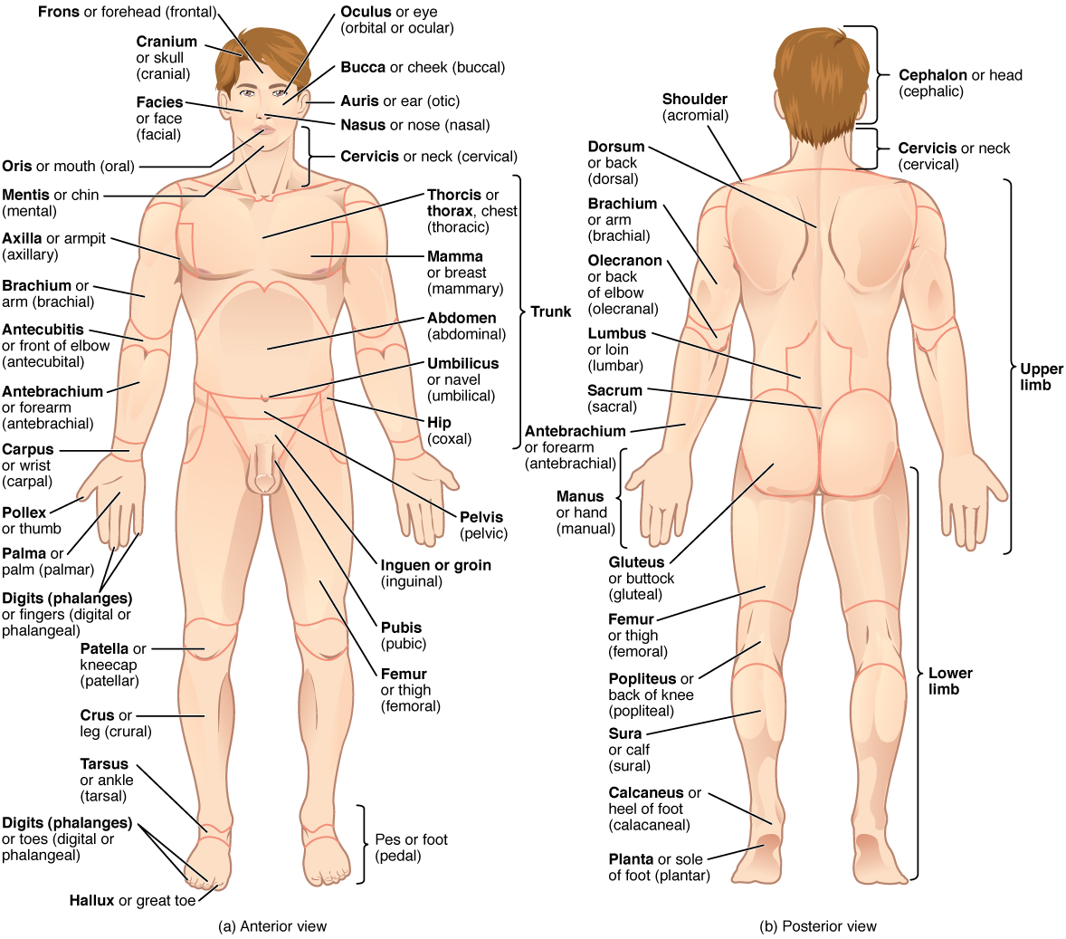
The human body’s numerous regions have specific terms to help increase precision. Figure \(\PageIndex\) has labeled each region with the correct anatomical term (seen in bold), while also listing the common name of the region. Additionally, the adjective form of the regional term is listed in parenthesis. Keep in mind that in the study of human anatomy, it is the bold terms listed in the figure that are used. Notice that the term “brachium” is reserved for the “upper arm” and “antebrachium” is used rather than “lower arm.” Similarly, “femur” designates the upper portion of the lower limb and “crus” is reserved for the portion of the lower limb between the knee and the ankle. You will be able to describe the body’s regions using the terms from the figure.
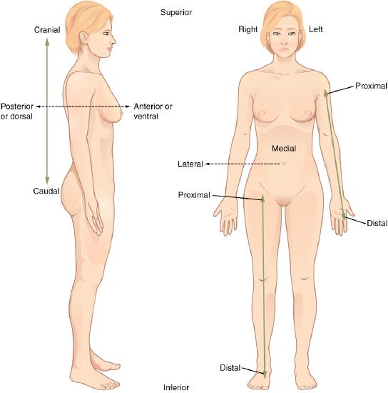
Certain directional anatomical terms appear throughout this and any other anatomy textbook (Figure \(\PageIndex\)). These terms are essential for describing the relative locations of different body structures. For instance, an anatomist might describe one band of tissue as “inferior to” another or a physician might describe a tumor as “superficial to” a deeper body structure. Commit these terms to memory to avoid confusion when you are studying or describing the locations of particular body parts.
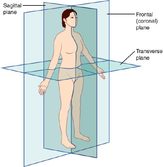
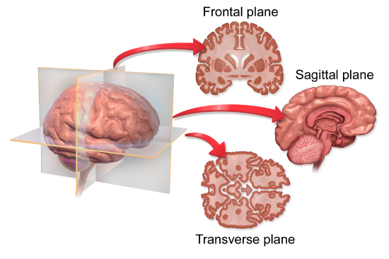
A section is a two-dimensional surface of a three-dimensional structure that has been cut. Modern medical imaging devices enable clinicians to obtain “virtual sections” of living bodies. We call these scans. Body sections and scans can be correctly interpreted, however, only if the viewer understands the plane along which the section was made. A plane is an imaginary two-dimensional surface that passes through the body. There are three planes commonly referred to in anatomy and medicine, as illustrated in Figure \(\PageIndex\).
The body maintains its internal organization by means of membranes, sheaths, and other structures that separate compartments. The dorsal (posterior) cavity and the ventral (anterior) cavity are the largest body compartments (Figure \(\PageIndex\)). These cavities contain and protect delicate internal organs, and the ventral cavity allows for significant changes in the size and shape of the organs as they perform their functions. The lungs, heart, stomach, and intestines, for example, can expand and contract without distorting other tissues or disrupting the activity of nearby organs.
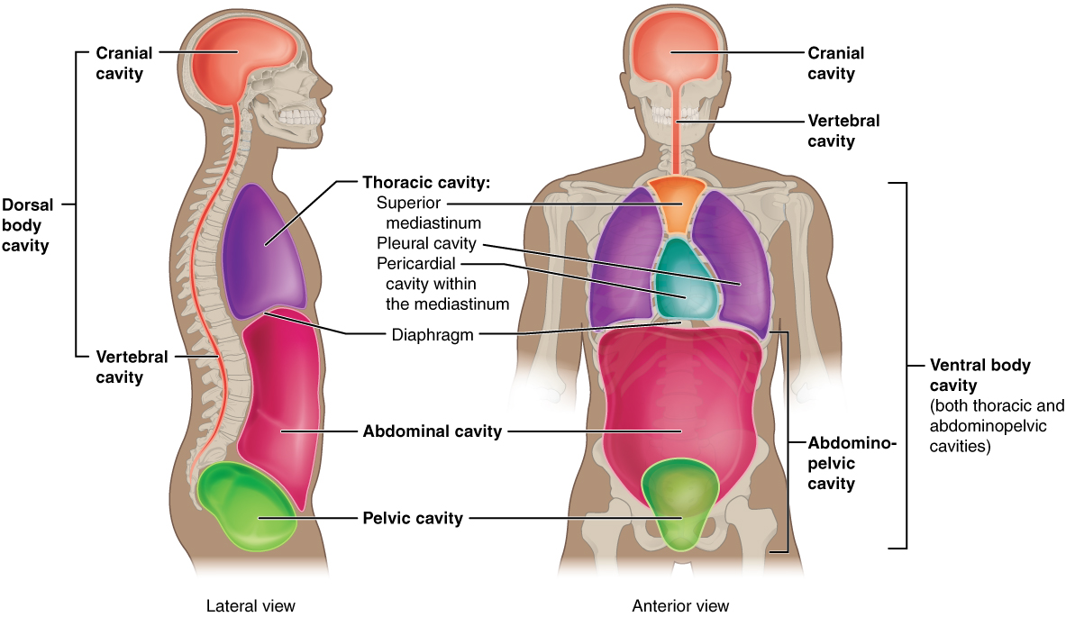
The posterior (dorsal) and anterior (ventral) cavities are each subdivided into smaller cavities. In the posterior (dorsal) cavity, the cranial cavity houses the brain, and the spinal cavity (or vertebral cavity) encloses the spinal cord. Just as the brain and spinal cord make up a continuous, uninterrupted structure, the cranial and spinal cavities that house them are also continuous. The brain and spinal cord are protected by the bones of the skull and vertebral column and by cerebrospinal fluid, a colorless fluid produced by the brain, which cushions the brain and spinal cord within the posterior (dorsal) cavity.
The anterior (ventral) cavity has two main subdivisions: the thoracic cavity and the abdominopelvic cavity (see Figure \(\PageIndex\)). The thoracic cavity is the more superior subdivision of the anterior cavity, and it is enclosed by the rib cage. It contains the pleural cavities, which house the lungs, and the mediastinum, the space between the lungs in the thoracic cavity. The mediastinum has a superior portion, which contains the trachea and esophagus, and an inferior portion containing the pericardial cavity, which surrounds the heart. The diaphragm forms the floor of the thoracic cavity and separates it from the more inferior abdominopelvic cavity. The abdominopelvic cavity is the largest cavity in the body. Although no membrane physically divides the abdominopelvic cavity into more specific compartments, it can be useful to distinguish between the abdominal cavity, the division that houses the digestive organs, and the pelvic cavity, the division that houses the organs of reproduction.
To promote clear communication, for instance about the location of a patient’s abdominal pain or a suspicious mass, health care providers typically divide up the cavity into either nine regions or four quadrants (Figure \(\PageIndex\)).
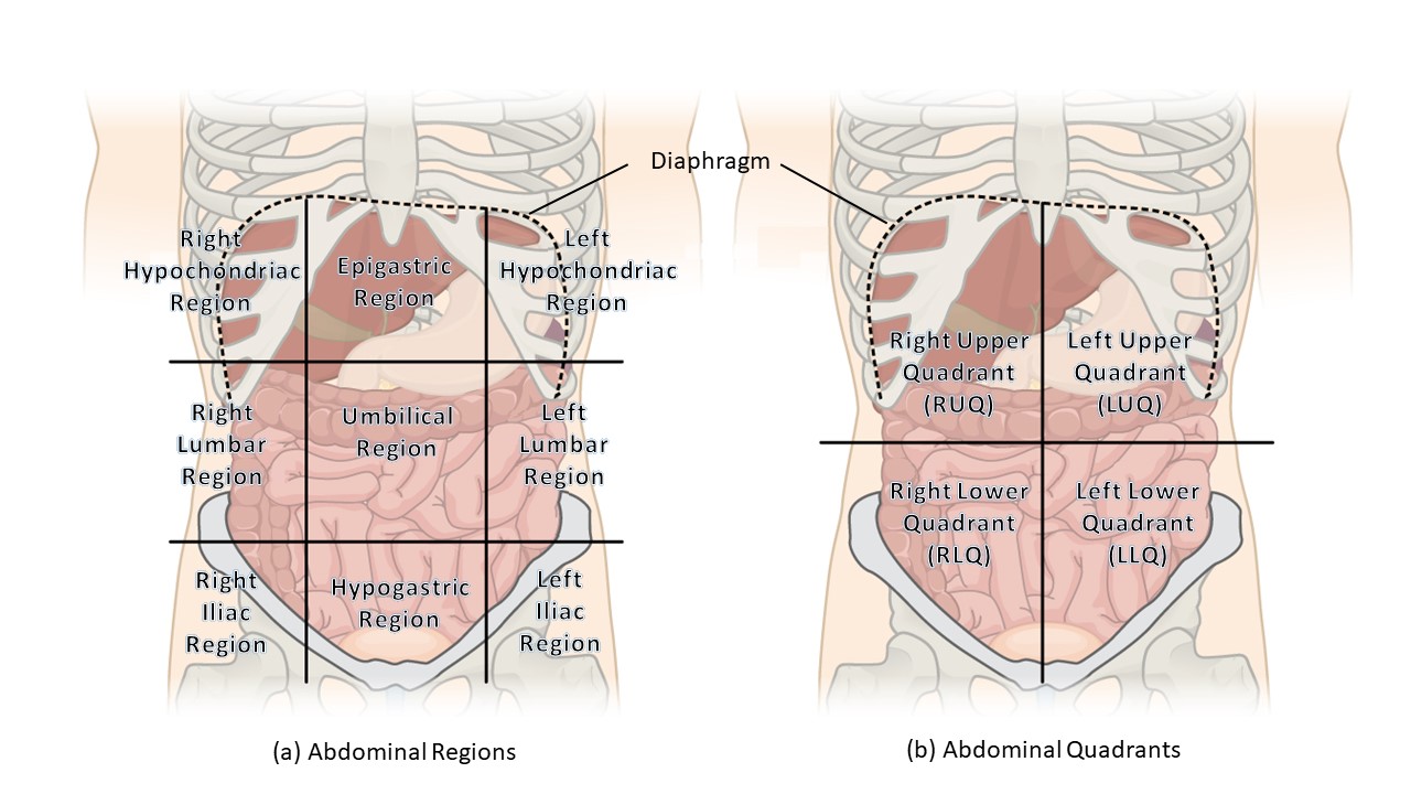
The more detailed regional approach subdivides the cavity with one horizontal line immediately inferior to the ribs and one immediately superior to the pelvis, and two vertical lines drawn as if dropped from the midpoint of each clavicle (collarbone). There are nine resulting regions:
The simpler quadrants approach, which is more commonly used in medicine, subdivides the cavity with one horizontal and one vertical line that intersect at the patient’s umbilicus (navel). This results in four quadrants:
A serous membrane (also referred to a serosa) is one of the thin membranes that cover the walls and organs in the thoracic and abdominopelvic cavities. The parietal layers of the membranes line the walls of the body cavity (pariet- refers to a cavity wall). The visceral layer of the membrane covers the organs (the viscera). Between the parietal and visceral layers is a very thin, fluid-filled serous space, or cavity (Figure \(\PageIndex\)).
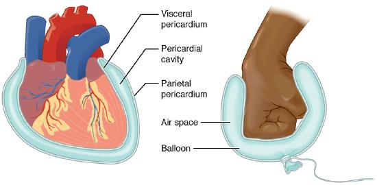
There are three serous cavities and their associated membranes. The pleura is the serous membrane that surrounds the lungs in the pleural cavity; the pericardium is the serous membrane that surrounds the heart in the pericardial cavity; and the peritoneum is the serous membrane that surrounds several organs in the abdominopelvic cavity. The serous fluid produced by the serous membranes reduces friction between the walls of the cavities and the internal organs when they move, such as when the lungs inflate or the heart beats. Both the parietal and visceral serosa secrete the thin, slippery serous fluid that prevents friction when an organ slides past the walls of a cavity. In the pleural cavities, pleural fluid prevents friction between the lungs and the walls of the cavity. In the pericardial sac, pericardial fluid prevents friction between the heart and the walls of the pericardial sac. And in the peritoneal cavity, peritoneal fluid prevents friction between abdominal and pelvic organs and the wall of the cavity. The serous membranes therefore provide additional protection to the viscera they enclose by reducing friction that could lead to inflammation of the organs.
A standard reference position for mapping the body’s structures is the normal anatomical position. Regions of the body are identified using terms such as “occipital” that are more precise than common words and phrases such as “the back of the head.” Directional terms such as anterior and posterior are essential for accurately describing the relative locations of body structures. Images of the body’s interior commonly align along one of three planes: the sagittal, frontal, or transverse. The body’s organs are organized in one of two main cavities — dorsal and ventral — which are further sub-divided according to the structures present in each area. The serous membranes have two layers — parietal and visceral — surrounding a fluid filled space. Serous membranes cover the lungs (pleural serosa), heart (pericardial serosa), and some abdominopelvic organs (peritoneal serosa).
Q. What is the position of the body when it is in the “normal anatomical position?”
A. The person is prone with upper limbs, including palms, touching sides and lower limbs touching at sides.
B. The person is standing facing the observer, with upper limbs extended out at a ninety-degree angle from the torso and lower limbs in a wide stance with feet pointing laterally
C. The person is supine with upper limbs, including palms, touching sides and lower limbs touching at sides.
D. None of the above
Answer
Q. To make a banana split, you halve a banana into two long, thin, right and left sides along the ________.
A. coronal plane
B. longitudinal plane
C. midsagittal plane
D. transverse plane
Answer
Q. The epigastric region is ________.
A. inferior to the hypogastric region
B. inferior to the right hypochondriac region
C. superior to the hypogastric region
D. superior to the left hypochondriac region
Answer
Q. The heart is within the ________.
A. cranial cavity
C. posterior (dorsal) cavity
D. All of the above
Answer
Q. If a bullet were to penetrate a lung, which three anterior thoracic body cavities would it enter, and which layer of the serous membrane would it encounter first?
Answer
A. The bullet would enter the ventral, thoracic, and pleural cavities, and it would encounter the parietal layer of serous membrane first.
abdominopelvic cavity division of the anterior (ventral) cavity that houses the abdominal and pelvic viscera anatomical position standard reference position used for describing locations and directions on the human body anterior describes the front or direction toward the front of the body; also referred to as ventral anterior cavity larger body cavity located anterior to the posterior (dorsal) body cavity; includes the serous membrane-lined pleural cavities for the lungs, pericardial cavity for the heart, and peritoneal cavity for the abdominal and pelvic organs; also referred to as ventral cavity caudal describes a position below or lower than another part of the body proper; near or toward the tail (in humans, the coccyx, or lowest part of the spinal column); also referred to as inferior cranial describes a position above or higher than another part of the body proper; also referred to as superior cranial cavity division of the posterior (dorsal) cavity that houses the brain deep describes a position farther from the surface of the body distal describes a position farther from the point of attachment or the trunk of the body dorsal describes the back or direction toward the back of the body; also referred to as posterior dorsal cavity posterior body cavity that houses the brain and spinal cord; also referred to the posterior body cavity frontal plane two-dimensional, vertical plane that divides the body or organ into anterior and posterior portions inferior describes a position below or lower than another part of the body proper; near or toward the tail (in humans, the coccyx, or lowest part of the spinal column); also referred to as caudal lateral describes the side or direction toward the side of the body medial describes the middle or direction toward the middle of the body pericardium sac that encloses the heart peritoneum serous membrane that lines the abdominopelvic cavity and covers the organs found there plane imaginary two-dimensional surface that passes through the body pleura serous membrane that lines the pleural cavity and covers the lungs posterior describes the back or direction toward the back of the body; also referred to as dorsal posterior cavity posterior body cavity that houses the brain and spinal cord; also referred to as dorsal cavity prone face down proximal describes a position nearer to the point of attachment or the trunk of the body sagittal plane two-dimensional, vertical plane that divides the body or organ into right and left sides section in anatomy, a single flat surface of a three-dimensional structure that has been cut through serous membrane membrane that covers organs and reduces friction; also referred to as serosa serosa membrane that covers organs and reduces friction; also referred to as serous membrane spinal cavity division of the dorsal cavity that houses the spinal cord; also referred to as vertebral cavity superficial describes a position nearer to the surface of the body superior describes a position above or higher than another part of the body proper; also referred to as cranial supine face up thoracic cavity division of the anterior (ventral) cavity that houses the heart, lungs, esophagus, and trachea transverse plane two-dimensional, horizontal plane that divides the body or organ into superior and inferior portions ventral describes the front or direction toward the front of the body; also referred to as anterior ventral cavity larger body cavity located anterior to the posterior (dorsal) body cavity; includes the serous membrane-lined pleural cavities for the lungs, pericardial cavity for the heart, and peritoneal cavity for the abdominal and pelvic organs; also referred to as anterior body cavity